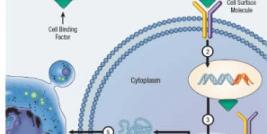
The term transfection is commonly used to describe the process of adding DNA into cells with a view to mediating protein expression of the transgene of interest. Although DNA transfection is the most commonly adopted technique, the term can also be applied to RNA transfection, where the purpose may also be to mediate protein expression or to achieve knockdown of the transcription of a specific gene, either by RNAi, ribozyme or antisense methodologies.
Transfection of Plasmid DNA
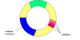
To obtain efficient gene transfer by non-viral means, plasmid DNA can be complexed by physico-chemical means to a variety of compounds in order to mediate efficient delivery into the cell's nucleus and then mediate subsequent protein expression of the gene of interest. Plasmids are small circular DNA molecules that naturally occur in bacteria, and are actually used by the bacteria to transfer genetic information non-genomically (i.e. without the need to alter the bacteria's own genome). Molecular Biologists have been able to harness this means of gene transfer in order to manipulate mammalian genes in a variety of ways (see section on genetic engineering). Indeed, plasmids can be considered the workhorses of genetic engineering as they are employed in almost all applications of molecular biology (see figure).
The plasmid DNA molecular comprises three basic features:
Origin of Replication: This allows the plasmid to replicate inside its bacterial host as the bacteria itself replicates. This is important as in order to make large quantities of DNA the plasmids must be 'grown' inside their bacterial hosts. After propagating the bacteria containing the plasmid in a special growth cocktail; the plasmid DNA can be extracted from the bacteria and purified for further manipulation (see section on genetic engineering).
Antibiotic Resistance: In order to select for the bacteria that have the plasmid DNA of interest, each copy of the plasmid must encode for an antibiotic resistance gene, e.g. genes encoding resistance to the antibiotics tetracyclin, kanamycin, streptomycin. These antibiotics can be added to the growth cocktail so that only the bacteria that contain the plasmid are able to grow.
Polylinker (or Multiple Cloning Site): A polylinker is important so that the gene of interest in the context of a eukaryotic protein expression cassette can be inserted into the plasmid (see section on protein expression). Plasmids can be treated with special enzyme proteins that cut the DNA at specific DNA sequences. The polylinker comprises many of these sequences that can be cut with a variety of different enzymes, with the function of inserting mammalian DNA into the plasmid (see section on genetic engineering).
Transfection Reagents
There are a large variety of transfection reagents that can be used to assist in the delivery of plasmid DNA into eukaryotic cell lines or primary cell cultures. Typically, the most efficient means of delivery, associated with lowest toxicity are lipid-based, such as liposomal, lipofectin, lipofectamine, etc. Below we describe some of the most frequently used transfection reagents often used to achieve efficient protein expression:
Calcium Phosphate Transfection
Calcium phosphate transfection, first performed in the early 1960s, was refined and systematically improved to result in a standard protocol which has changed little since the early 1970s. In many cases it remains the system of choice for transferring plasmid DNA into a variety of cell cultures and packaging cell lines. It is particularly important in the production of recombinant viral vectors.
The technique relies on precipitates of plasmid DNA formed by its interaction with calcium ions. It is a very inexpensive and simple technique to perform. Plasmid DNA is mixed in a solution of calcium chloride, and then is added to a phosphate- buffered solution. Over a period of 20 minutes a fine precipitate forms in the solution, and this solution is then added directly to the cells in culture. Transfer efficiency, the number of cells which express the desired gene, is usually quite limited and only reaches levels greater than 10% in a few specific cell lines. In many cases, the level is less than 1%. Transfection efficiencies can be improved in some cell lines by 'shocking' the cells with DMSO or glycerol. Cells can be either transiently or stably transfected using this technique. Levels of stable transfectants can be improved using bis-hydroxyethylaminoethansulfonate (BES). The DNA precipitates are thought to enter the cell by endocytosis. Although this technique has minimal cellular toxicity, and is both simple and inexpensive, the low level of transgene expression prompted development of other techniques. Little or no transgene expression has been observed in vivo following calcium phosphate transfection.
DEAE-dextran is another compound that interacts and delivers DNA into cells. It is more reproducible than calcium phosphate transfection but only works in a select few cell lines. Also, DEAE-dextran-mediated gene transfer results only in transient transfections. The exact mechanism of DNA uptake mediated by this technique is not understood.
Lipid-Based Transfection
Lipid transfection is referred to with a variety of names: lipid-mediated gene delivery, lipofection, and liposome-based gene transfection, among others. Each name refer to the same process, though: using lipids to cause a cell to absorb DNA from outside itself.
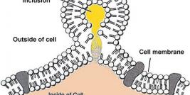
The mechanism of cationic lipid-mediated transfection originates with the basic structure of cationic lipids, a positively charged head group and one or two hydrocarbon chains. The positive surface charge of the liposomes mediates the interaction of the nucleic acid and the cell membrane, allowing for fusion of the liposome/nucleic acid (“transfection complex”) with the negatively charged cell membrane. The transfection complex is thought to enter the cell through endocytosis. Once inside the cell, the complex must escape the endosomal pathway and diffuse through the cytoplasm to the nucleus in order to mediate protein expression.
Electroporation Systems
Electroporation is the application of high voltage to a mixture of DNA and cells in suspension. The cell-DNA suspension is placed between two electrodes and subjected to an electrical pulse. The DNA enters the cells through holes formed in the cellular membrane during the electrical pulse. The DNA is trapped within the cytoplasm upon termination of the electrical pulse. For efficient gene transfer, electroporation depends upon the nature of the electrical pulse, the distance between the electrodes, the ionic strength of the suspension buffer, and the nature of the cells. Best results have often been obtained from rapidly proliferating cells. Electroporation of mammalian cells is an inefficient technique since many cells do not survive the high voltage nature of this procedure. Because of its unique characteristics, electroporation has been difficult to properly design for in vivo applications.
Micro-Injection of DNA
Microinjection delivers plasmid DNA directly into the cell's nucleus. Using the light microscope, a glass pipette is guided into the nucleus and a small amount of DNA or RNA injected. In doing so, both cytoplasmic and lysosomal degradation of the injected material is avoided and efficient gene expression can be expected from the surviving cells. Unfortunately, this technique is extremely labor intensive and requires well isolated cells.
Since safety and efficiency can be closely monitored using microinjection, it may one day prove useful in germline gene therapy applications. Microinjection of a particular gene into a cell at a specific time during development has the potential to alter the genotype and phenotype of every cell formed thereafter. Germline modification has already been accomplished in many mammals, which have been termed transgenic animals, ranging in size from mice to cattle. Germline modifications in humans for the correction of specific disorders is far from a present reality and is fraught with ethical considerations.

