Article by: Lilja E. Laatikainen (1) and Mikko O. Laukkanen (2)
Experimental hind limb injury is widely used to study the effect of transgenes, stem cells, or pharmaceuticals on e.g. new vessel formation (1,2,3,4). The degree of the injury depends on the number and the location of the vessels ligated, therefore having an impact on the recovery process and the monitoring of the transgene response. Even though terminally differentiated myofibers are not able to proliferate the skeletal muscle has a remarkable regenerative potential, which is thought to be due to the satellite cell differentiation as response to injury (5,6). Selection of the species between mouse, rat, and rabbit depends on the purpose of the study and has an impact on the method of vessel ligation. In the current review we focus on to describe the hind limb injury execution in a rat model.
Reagents needed
Fentanyl-fluanisone (Hypnorm), Midazolame (Dormicum), Sterile water, Naloxone, 2-methylbutane cooled with liquid nitrogen
Instruments
Surgery microscope, Surgery light, Warming lamp, Microsurgery instrument set, 1 ml syringes, Glass tuberculin syringe, 0.25 ml, 30G needles, Mersilk suture, U.S.P. size 6-0, Vicryl absorbable suture, U.S.P. size 5-0, with 11mm 3/8c curved needle, Shaving instrument, Disinfectant
PROTOCOL
Anesthesia
As anesthetics we have used a mixture of fentanyl-fluanisone (trade name Hypnorm; Janssen Pharmaceutica, Beerse, Belgium) and midazolame (trade name Dormicum; Roche, Basel, Switzerland) because of their reliability and safety for the animals. The two anesthetics should first be diluted in water 1:1 and then combined resulting in the final dilution of 1:1:2 (Hypnorm:Dormicum:water). The solution is administered by a single intraperitoneal (i.p.) 100µl injection for an animal in the weight range of 60-90g, 150µl for animals of 90-130g, and 200µl for animals of 130-160g. This should provide anesthesia for approximately 30-40 minutes that, however, must be monitored continuously during the surgical procedure by e.g. pinching the skin flap between toes with forceps. An additional dose (30-100µl) of anesthetic solution should be estimated according to the size of the animal and the remaining time of the procedure. After the wound is properly sutured and disinfected we have injected 50µl of naloxone i.p. to reverse the anesthesia. The animal is then placed under a heat lamp until it is awake.
Vessel ligation
For the microsurgery a proper working environment and equipments are needed, such as operating microscope with external light source for adequate lighting of the area. After the ventral side of the hind limb of the anesthetized the animal is shaved the skin is incised with small scissors following the line along the femur. However, the incision should not be longer than 15mm starting from the inguinal region to enable proper healing since adhesions often develop along the wound. The femoral artery is ligated at two sites: 1) distally just above the popliteal and saphena artery bifurcation point and 2) proximally close to circumflexa femoralis lateralis branch. In addition, the third ligation is made to close circumflexa femoralis lateralis to prevent blood flow to ischemic region between two femoral artery ligations. Perma-Hand Mersilk suture, U.S.P. size 6-0 (Johnson&Johnson Intl, St-Stevens-Woluwe, Belgium) cut into approximately 10-15mm pieces is a good choice for vessel ligation. The proximal femoral artery and the circumflexa femoralis lateralis are situated deep in the inguinal region usually covered under a layer of adipose tissue, which should be damaged as little as possible. Collateral damage should be kept to a minimum to avoid overt inflammation of the tissue that may interfere with the intended ischemic inflammation.
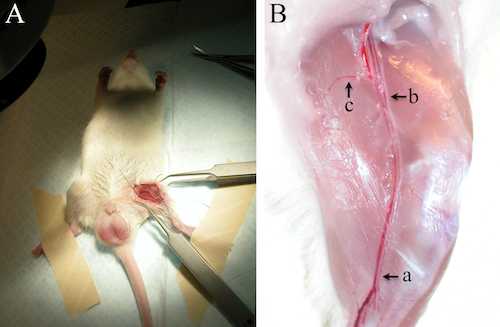
Figure. Rat hind limb injury. A. The vessels for the ligation covered only by a few transparent layers of connective tissues are visible above the muscles. Attention is needed not to damage the neurons located immediately under the femoral artery. B. Ligated vessels shown post-mortem. In our model we have ligated the following three vessels; a. femoral artery above popliteal and saphena artery bifurcation point, b. femoral artery close to circumflexa femoralis lateralis branch, c. circumflexa femoralis lateralis
Gene transfer and tissue isolation
We have injected the virus vector into the muscle in 50µl volume (5x10µl doses) to avoid unwanted stretching and damage to the tissue that cause unnecessary inflammation. Serial injections enable even distribution of the virus when the injection sites are a few millimeters apart. However, due to the compact structure of the muscle, the infected area will stay patchy. We have used a 0.25 ml glass tuberculin syringe (Sigma, Saint Louis, MI, USA) to achieve adequate accuracy and short 30G needles to minimize tearing of the tissue. In our experience, it is best to slowly pull out the needle while injecting the virus vector and to pull out the tip of the needle only after a few seconds when pressure is no more applied on the syringe; this prevents most of the leakage from the channel.
The wound is closed with coated Vicryl absorbable suture, U.S.P. size 5-0 with 11mm 3/8c curved needle (Johnson&Johnson Intl, St-Stevens-Woluwe, Belgium). Absorbable suture has the advantage of disappearing without the need to sedate the animals for removal and thus they can be followed uninterrupted during follow-up period.
Due to the regenerative potential of the rat skeletal muscle the animals on regular basis are able to use their ligated limbs normally already in 1-2 days. Some hemorrhage at metatarsus could be visible initially that should disappear within a few days. Based on the hematoxylin-eosin staining the injury is healed almost completely in two weeks, which however, depends on the severity of the damage. In general, it is recommended that only one person is executing the injury in the animal series since the differential working methods between researchers are crucially affecting the degree of the injury and therefore also the reliability of the data.
At the end of the follow-up period the samples are collected around gene transfer sites for expression analysis and histological staining. Since the muscle tissue is sensitive to artifacts caused by the freezing method we have prepared the histological samples for cryo sectioning by freezing a neatly cut portion of thigh muscles in pre-frozen 2-methylbutane, which is kept in a metal container in liquid nitrogen until it begins to solidify. The tissue should be kept in the freezing solution at least for 30 seconds and maintained in low temperatures until sectioning. The thickness of the cryo sections should be approximately 8-10 μm. In case the freezing artifacts (a large vacuole like formations in the tissue section) are visible they can be avoided by cutting thicker 30 μm sections.
1University of Turku, Medicity Research Laboratory, 20520 Turku, Finland, 2Fondazione IRCCS SDN, 80143 Naples, Italy

