Article by: Min Song*
What is HIV-1?
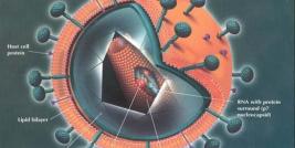
Human immunodeficiency virus (HIV) is the causative agent of acquired immunodeficiency syndrome (AIDS) [1]. According to the report from World Health Organization (WHO), in 2009, there were 33.3 million people living with HIV/AIDS, with 2.6 million new infections and 1.8 million deaths due to AIDS (http://www.who.int/hiv/data/2009_global_summary.png). HIV infection affects a large area and spreads actively ever since it was identified, with a significant number of AIDS deaths occurring in Sub-Saharan Africa [Greener R (2002) AIDS and macroeconomic impact. In S, Forsyth (ed) State of the Art: AIDS and Economics IAEN: pp. 49-55].
HIV infects essential cells present in the human immune system, including CD4+ T cells, macrophages, etc. Once the number of CD4+ T cells decreases to a critical level, the cell-mediated immunity is destroyed, rendering the infected individual more susceptible to opportunistic infections or cancer [3]. Two types of HIV viruses have been characterized: HIV-1 and HIV-2. HIV-1 is more virulent and infective, accounting for the majority of HIV infections worldwide [4].
HIV-1 life cycle
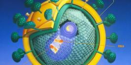
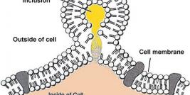
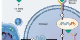
Understanding the nature of HIV-1 replication is essential for identifying intervention targets to treat HIV-1 infection. Macrophages and CD4+ T-lymphocytes are two important targets of HIV-1. HIV-1 enters host cells by the adsorption of glycoproteins on its surface to receptors on the target cells. The initial binding is followed by the fusion of viral envelope with the cell membrane and the release of the content of HIV-1 particle, HIV-1 capsid, into the host cells [5,6,Coffin JM (1990) Retroviridae and their replication. In Virology (Fields, BN and Knipe, DM eds), 2nd edit: pp. 645-708, Raven Press, NY]. Shortly after the viral entry, a viral enzyme named reverse transcriptase converts the single stranded viral RNA genome into double-stranded viral DNA via revere transcription [Coffin JM (1990) Retroviridae and their replication. In Virology (Fields, BN and Knipe, DM eds), 2nd edit: pp. 645-708, Raven Press, NY]. After reverse transcription, the viral DNA is imported into the cell nucleus. Another viral enzyme, integrase, integrates the viral DNA into the host chromosome, generating HIV-1 provirus [Coffin JM (1990) Retroviridae and their replication. In Virology (Fields, BN and Knipe, DM eds), 2nd edit: pp. 645-708, Raven Press, NY]. Once integrated, the virus can either lie dormant in host cells, or begin the production of new viral RNA and proteins. During active viral replication, the integrated provirus servers as the template for the synthesis of viral mRNAs. These mRNAs are exported into the cytoplasm, where they are translated into structural, regulatory, and accessory proteins that are essential for viral replication [Coffin JM (1990) Retroviridae and their replication. In Virology (Fields, BN and Knipe, DM eds), 2nd edit: pp. 645-708, Raven Press, NY]. Following the synthesis of the viral proteins and posttranslational modification, the virus assembly takes place at the plasma membrane of the infected cell. Viral RNAs and proteins are packaged to form virus particles, which will then bud off from the host cells. Maturation, which can take place either during budding or in the immature virion after it is released from the host cell, turns the virion into an infectious particle that is ready to infect another cell and initiate the replication cycle all over again [Coffin JM (1990) Retroviridae and their replication. In Virology (Fields, BN and Knipe, DM eds), 2nd edit: pp. 645-708, Raven Press, NY].
Gene therapy of HIV-1 treatment
The development of highly active anti-retroviral therapy (HAART) has substantially reduced the mortality among infected individuals of HIV [8]. Although HAART regimens have been improved quite significantly, cost, side-effects, and the development of multidrug resistance have raised the concerns about the effectiveness of HAART [9,10]. Protein-based and RNA-based anti-HIV-1 strategies have been widely used as alternative methods for anti-HIV-1 gene therapy [Zaia JA, Cairns, J.S., and Rossi, J.J. (2004) AIDS and hematopoietic cell transplantation: HIV infection, AIDS lymphoma, and gene therapy. In Thomas' hematopoietic cell transplantation, 3rd ed (KG Blume, SJ Forman, FR Appelbaum Eds Blackwell Sci, Boston,12].
Protein-based gene therapy strategies include transdominant negative mutants, toxins, antibodies, etc [Zaia JA, Cairns, J.S., and Rossi, J.J. (2004) AIDS and hematopoietic cell transplantation: HIV infection, AIDS lymphoma, and gene therapy. In Thomas' hematopoietic cell transplantation, 3rd ed (KG Blume, SJ Forman, FR Appelbaum Eds Blackwell Sci, Boston,12]. CXCR4 and CCR5 are two important co-receptors for HIV-1 infection. Therefore, intracellular expression of SDF-1, the ligand for CXCR4, or RANTES, the ligand for CCR5, may decrease the number of functional co-receptors available to participate in viral entry and confer resistance to the viral infection to a certain degree [13,14]. Viral Rev protein is important for productive viral replication. A mutant form of Rev, Rev M10, has been reported to compete with wild-type Rev to bind to Rev-responsive element. Expression of Rev M10 prolonged the survival of CD4+ cells [15]. These results collectively suggest the great potential of protein-based methods in the of HIV infection.
RNA-based gene therapy approaches include the usage of antisense RNAs molecules, RNA decoys, or the method of RNA interference (RNAi), etc [Zaia JA, Cairns, J.S., and Rossi, J.J. (2004) AIDS and hematopoietic cell transplantation: HIV infection, AIDS lymphoma, and gene therapy. In Thomas' hematopoietic cell transplantation, 3rd ed (KG Blume, SJ Forman, FR Appelbaum Eds Blackwell Sci, Boston]. It was observed in an in vitro model that antisense oligonucletides could inhibit the replication of HIV-1, and the antiviral activity is associated with the target to which the oligonucleotide is complementary [16,17]. RNA decoys are molecules homologues to wild-type ligands. Therefore, they will compete with wild-type ligands to bind viral proteins and interfere with viral replication. For instance, Tat activation-response region (TAR) decoys have been used to inhibit the interaction between viral protein Tat and natural transcriptional promoter of HIV-1 [18]. The strategy of RNAi has been adopted and utilized in a wide range of different researches including the treatment of HIV-1. It was reported that the usage of synthetic siRNAs or plasmid-derived siRNAs interfered with HIV-1 replication by degrading the viral genomic RNA [19]. Together, these reports demonstrate the efficacy of RNA-based strategies in modulating the replication of HIV-1.
Animal models for gene therapy of HIV-1
Animal models that closely reflect human physiology and pathology are extremely valuable for the study of human diseases as well as preclinical testing of novel drugs. For the research on human HIV-1 infection, severe combined immunodeficiency (SCID) mice have been utilized as the base from which two important rodent models were established. The hu-PBL-SCID chimeras are generated by the injection of healthy human peripheral blood lymphocytes (PBLs) into SCID mice [20], while the thy/liv SCID-hu chimeras are generated by implanting human fetal liver and fetal thymus tissue into SCID mice [21].
The hu-PBL-SCID mouse model has been applied to a number of studies on HIV pathogenesis, vaccines, etc. The usage of this model, however, is limited. T cells become anergic in hu-PBL-SCID mice progressively. Also, human lymphocytes in these mice are functional only within the first two to three weeks following PBL injection [20]. To evade the weakness of this model, a new hu-PBL-SCID mouse model, which could support persistent and productive HIV replication, was developed and utilized in a gene therapy study. Beta interferon contains anti-HIV-1 activities. Transfer of human CD4+ T cells containing the beta interferon retroviral vector significantly reduced the HIV-1 replication while facilitated the proliferation of human CD4+ T cells in HIV-1 infected hu-PBL-SCID mice [22]. These results suggest the importance of hu-PBL-SCID mouse model in gene therapy studies of HIV-1.
The thy/liv SCID-hu mouse model was initially designed based on the hope that the mouse recipient would allow for the growth of implanted human fetal liver, thymus, or lymph node tissue. The observation of long-term multilineage human hematopoiesis, including T lymphopoiesis, in thy/liv SCID-hu mice facilitated the application of this model to a broad range of investigative studies [21]. The SCID-hu mouse model was also developed to permit the transfer and reconstitution of hematopoietic stem cell. Human CD34+ hematopoietic progenitor cells obtained from different human organs will eventually develop into normal T lymphocytes after introduced into thy/liv SCID-hu mice. It was reported that retrovirus-transduced CD34+ cells could reconstitute the human thymus, leading to the generation of mature CD4+ and CD8+ lymphoid cells [23,24]. Taken Together, thy/liv SCID-hu mouse model provides an ideal in vivo system to study HIV-1 gene therapy.
Viral vectors for gene therapy of HIV-1
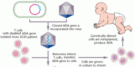
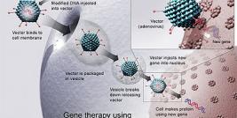
Viral based vectors for gene therapy are continually being studied, developed, and improved. The effectiveness of this technology is not only approved in research, but also demonstrated in clinical trails [25]. Vectors based on murine leukemia virus (MLV) are the first viral vectors applied to gene therapy and continue to serve as one of the most reliable means, however, lentivirus-based retroviral vectors are of particular interest for HIV-1 treatment, for two reasons. First, lentiviral vectors can transduce non-dividing cells. Secondly, compared to that of MLV, the lentiviral integration is less susceptible to alteration or any influences on gene expression [26,27].
Despite the advantages regarding the usage of lentivral vectors, there are safety concerns as well. The most prominent concern is the generation of replication-competent lentivirus due to viral recombination [28]. Therefore, other viruses are also evaluated as vectors for therapy of HIV-1, one of which is human foamy virus (HFV). HFV based vectors possess a number of advantages. Unlike MLV vectors, HFV vectors are not inactivated by human serum. The transduction rate of HFV vectors on stationary-phase cells is higher than that of MLV, although HFV preferentially transduce dividing cells. In addition, unlike some lentiviral vectors, HFV vectors are not targeted by anti-HIV sequences [29].
Another well recognized gene delivery vehicle is recombinant simian virus-40(rSV40)-derived vectors. These vectors are capable of infecting a wide variety of different cells that are currently targeted in HIV-1 therapy. They could be concentrated to high titers therefore the usage of these vectors to treat cell pools or large organs is relatively easier. Moreover, SV40 based vectors do not elicit strong immune responses that cause the elimination of infected cells. The effective delivery of anti-HIV-1 genes to CD34+ cells due to the high transduction efficiency of these rSV40-derived vectors has been well documented [30]. These results collectively suggest rSV40-derived vectors possess considerable promise in the gene therapy of HIV-1.
Conclusions
AIDS is one of the most destructive pandemics in recorded human history. Significant progresses in characterization of HIV-1 replication and antiretroviral treatment have been realized ever since the virus was initially identified. Gene therapy strategies to treat HIV-1 infection have been developed and improved. Further studies to improve the strategies of HIV-1 treatment are needed and still in progress.
*300 Pasteur Drive, Department of Genetics, Stanford University, Stanford, CA, 94305

