disease targets

Gene Therapy for Duchenne Muscular Dystrophy

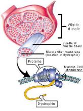
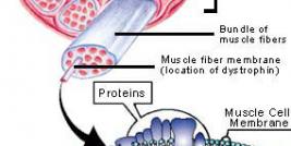
Duchenne muscular dystrophy (DMD) is an X-linked genetic disease characterized by the absence of dystrophin in the muscle. This large protein of 427 kDa is encoded by a 14 kb mRNA (79 exons) (1). This dystrophin protein is located under the membrane of the muscle fiber and interacts with other trans-membrane proteins. It is needed to insure mechanical stress resistance during muscle contraction. The lack of dystrophin weakens the sarcolemma and thus makes fibers less resistant to stress. When a fiber is damaged, the satellite cells (stem cells located near the muscle fibers) are activated and proliferate to allow fiber regeneration (2). In the case of a DMD patient, fibers are frequently damaged due to the lack of dystrophin. The regeneration is also ensured by the proliferation of satellite cells though their number decreases rapidly and when there are no more satellite cells, the average diameter and the number of fibers progressively decrease bringing the development of fibrosis and fat infiltrations into the muscles. There is currently no treatment for DMD and life expectancy is between 20 to 25 years of age.

Controlling Gene Expression in Ischemic Tissues

Prolonged ischemic insult can cause irreversible tissue injuries. Despite advances in medical and surgical therapies, myocardial infarction and stroke, both consequences of ischemic insults, remain to be the top two causes of morbidity and mortality in the Western world, with survivals usually carrying permanent disabilities. Treatments that help restoring blood flow to ischemic area remain to be one of the most important therapeutic goals. Enhancing the innate angiogenesis by exogenous delivery of angiogenic factors lessens the ischemic injury. However, uncontrolled angiogenic gene expression can cause some unwanted side effects. In this mini-review, we describe two systems that can be used to mediate hypoxia-inducible and tissue-specific gene expression.

p53 Gene Therapy

Replication-defective adenoviruses are widely used as gene transfer vectors to deliver cytostatic or tumour suppressor genes into a variety of cancers, yielding some of the most promising results in the clinic. Indeed, the most successful adenoviral vectors used to date are those that are designed to deliver p53 into tumours. Tumor cells have lost the function of p53 because of mutations in the DNA-binding region of the molecule, a feature that is present in over 50% of all human malignancies. A number of studies have demonstrated that transfer of wild type (wt) p53 gene is able to suppress tumor cell proliferation. Moreover, platinum-based chemotherapy enhances mutations in the p53 in the heterogenous cell population; transfer of the wild type p53 gene enhanced the sensitivity of chemoresistant cells to cisplatin and cisplatin-induced apoptosis (Kigawa et al, 2002). This has led to a significant amount of clinical trials where wt p53 has been re-introduced to tumour cells by the use of first-generation adenoviral vectors.