Article by: Jani Räty
Imaging of gene therapy treatments
Gene therapy has developed greatly and the amount of patients treated in various clinical trials has continued to increase during the last decade (with 89 approved trials 2007, http://www.wiley.co.uk/genmed/clinical/).
In addition to assessing the outcome of the treatment, non-invasive in vivo imaging has emerged as essential tool to:
- identify the fate of the vector in the tissue
- quantify the injected dose in the organs
- evaluate the toxicity
- analyze the gene expression pattern
- evaluate the therapeutic gene expression level
For a more comprehensive review see Raty et al. 2007b.
Ideally, on the path to clinical use, it would prove valuable to address all these points during the development of new and safe gene delivery vectors (Raty et al. 2008). In this article we give a brief overview of the imaging methods employed to evaluate dissemination of gene transfer vectors following clinical administration.
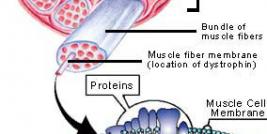
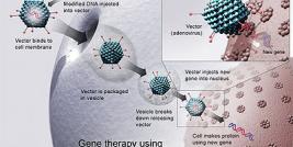
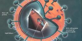
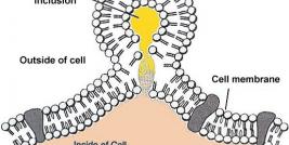
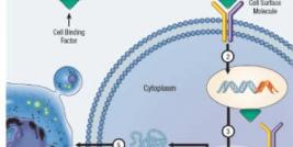
Transduction and biodistribution imaging
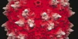
The current imaging methods in gene therapy can roughly be divided in two, namely transduction and biodistribution imaging. Transduction refers to use of viral or non-viral vectors to introduce a transgene to patient’s cells. While the transduction imaging visualizes the transgene- mediated protein production in the cells, the biodistribution imaging visualizes the actual systemic distribution of the gene delivery vectors (Kootstra and Verma 2003). This imaging methods enables to researcher to study the spreading of the vectors in subjects body, instead of waiting for the gene expression. In some cases, this might be vital since a virus may be unable to express the transgenes in all cells while being still able to accumulate the same cells and tissues. This could decrease the efficiency of the treatment and cause problems with the immunosystem of the patient. Therefore it is important to evaluate both the particle kinetics and transgene expression of viral vectors in vivo.
MRI
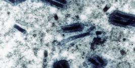
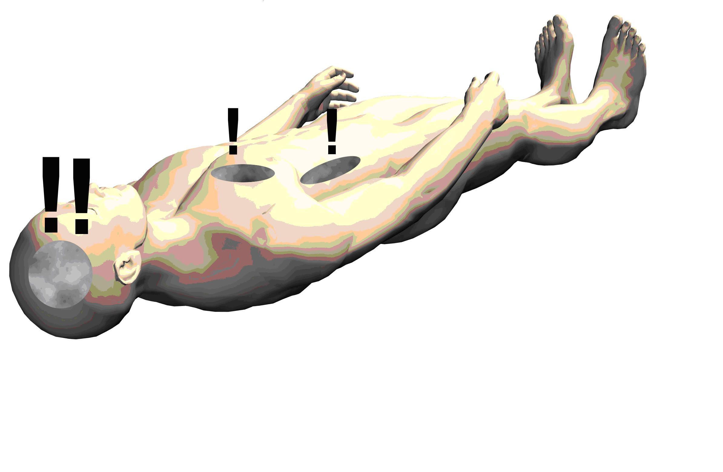
Imaging technologies allowing non-invasive visualization of the body are based on different forms of energy interacting with tissues. Methods such as magnetic resonance imaging rely on energy-tissue interactions and can be used with gene therapy with suitable contrast agents (Raty et al. 2006). MR- imaging is widely used on clinical imaging and it can detect signal in great details.
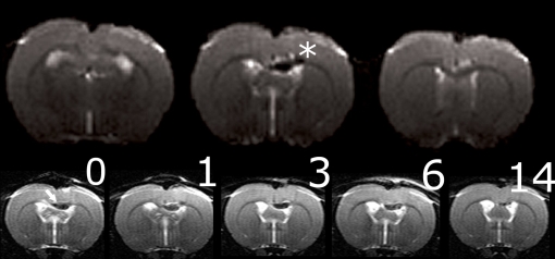
By MRI, viral particle accumulation to brain tissue can be detected and followed daily, if needed.
SPECT / PET imaging
Other important methods, such as single photon emission tomography (SPECT) and positron emission tomography (PET) require the administration of radioactive reportes probes (Raty et al. 2007a). With these methods, very low amounts of vectors can be detected and followed in real time.
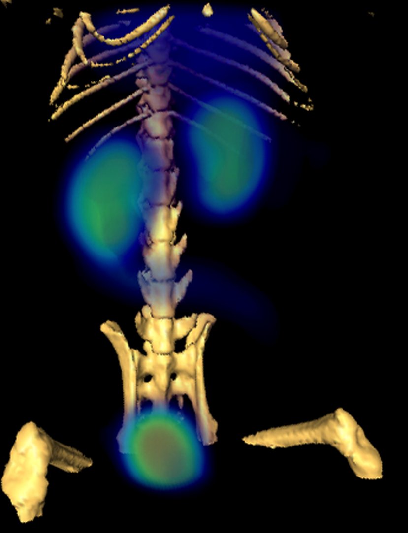
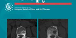
With SPECT-imaging, the biodistribution of the viral particles can be seen throughout the subject.
Optical imaging
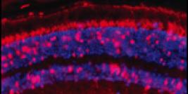
In addition, some techniques rely on the visible light, using fluorescence or luminescence methods to detect the viral particles or transgene expression (Smith et al. 2005). However, because light penetration through living tissue is not very efficient, these methods have a limited clinical use.
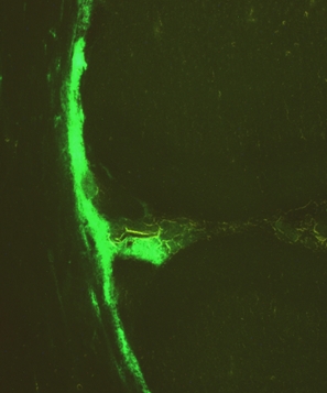
Optical imaging enables seeing transduced cells by using visible light wavelengths.
The future
Considering the emergence of molecular medicine through genomics research, therapeutic agents and specialized imaging techniques are becoming increasingly important (Bogdanov, Jr. 2003). When the gene therapy comes more common, the imaging of the administration safety and outcome of the therapy with routinely used imaging modalities is coming increasingly important.